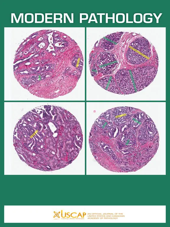
Historically, fibrolamellar carcinoma (FLC) has been diagnosed by analyzing the histology (microscopic anatomy) and morphology (shape, size and structure) of the tumor tissue. Frequently, that examination is combined with immunostaining for cytokeratin 7 and CD68. That immunostaining uses antibodies to see if proteins typically found in FLC are present. However, accurate diagnosis of fibrolamellar carcinoma continues to be a challenge.
This paper proposed a new method to confirm diagnosis of FLC – the presence of the DNAJB1-PRKACA gene fusion. The proposed technique is called break-apart fluorescent in situ hybridization (break-apart FISH). In this assay, a previously published break-apart probe set for the PRKACA locus was used. In it, a green and a red probe are used that target adjacent sequences of DNA on the PRKACA gene. The green probe binds to the PRKACA gene while the red probe binds to the adjacent sequence. In normal liver tissue, the green and red light emit from the same location, and a yellow light is seen. However in FLC, the 400 kb deletion results in the loss of the DNA covered by the red probe, so only a green light is seen in that location. This study tested using this FISH test in over 100 cases of FLC from North America, South America, and Europe.
The study concluded PRKACA FISH is a powerful tool to confirm the diagnosis of fibrolamellar carcinoma, especially in challenging cases. The authors propose that the best approach for diagnosing FLC is based on compatible morphology plus molecular confirmation (using a technique like PRKACA FISH), or if not available, then confirmation by CK7 and CD68 immunohistochemistry.
The complete text of the published article can be accessed here.
